In a groundbreaking development, a team of scientists at the University of Tokyo have unveiled a new and improved mid-infrared microscope that allows for the visualization of structures inside living bacteria at the nanometer scale. This remarkable achievement, featured in a recent publication in Nature Photonics, marks a significant advancement in the realm of microscopy, particularly in the field of biological research.
The Limitations of Traditional Microscopy Techniques
Traditionally, mid-infrared microscopy has been plagued by its limited resolution capabilities, especially when compared to other more advanced microscopy techniques. For instance, while super-resolution fluorescent microscopes can achieve resolutions down to tens of nanometers, mid-infrared microscopy has typically been restricted to around 3 microns. This discrepancy has hindered the widespread adoption of mid-infrared microscopy in biological research, as the low resolution has posed significant limitations in visualizing intricate details within live cells.
A 30-Fold Improvement in Resolution
However, the team at the University of Tokyo has managed to overcome this barrier by achieving a remarkable resolution of 120 nanometers with their newly developed mid-infrared microscope. This represents a 30-fold enhancement in resolution compared to conventional mid-infrared microscopes, enabling researchers to view samples with unprecedented clarity at a much smaller scale. This breakthrough has profound implications for various fields of research, including infectious diseases, where the ability to observe cellular structures at the nanometer level can provide valuable insights into disease mechanisms and treatment strategies.
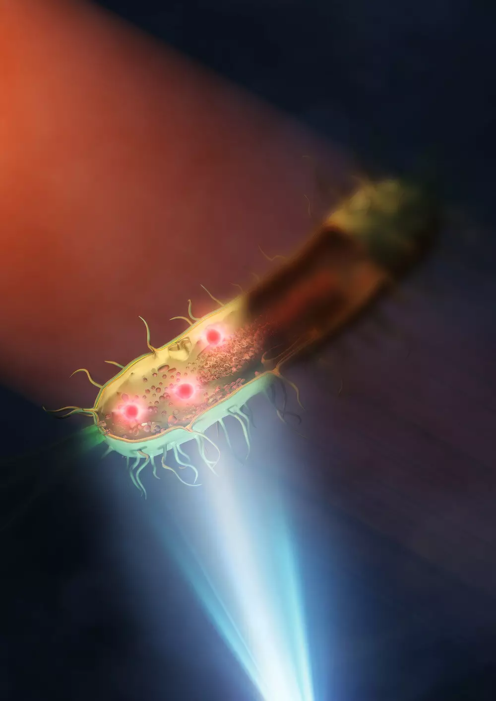
The key innovation behind this breakthrough lies in the utilization of a “synthetic aperture” technique, which involves combining multiple images captured from different illuminated angles to create a more detailed overall picture. By placing the sample – in this case, living bacteria such as E. coli and Rhodococcus jostii RHA1 – on a silicon plate that reflects visible light and transmits infrared light, the researchers were able to eliminate the need for two lenses and instead use a single lens for better illumination. This resulted in clearer and more precise imaging of the intracellular structures of bacteria, offering new avenues for studying antimicrobial resistance and other pressing global health issues.
Looking ahead, Professor Takuro Ideguchi from the Institute for Photon Science and Technology at the University of Tokyo expressed confidence in the continued improvement of this innovative technique. By exploring the use of better lenses and shorter wavelengths of visible light, the spatial resolution of the mid-infrared microscope could potentially be pushed below 100 nanometers, unlocking even greater detail in the visualization of live cells and biological processes. This represents a promising avenue for future research and discovery in the field of microscopy and life sciences.
The development of the enhanced mid-infrared microscope at the University of Tokyo stands as a remarkable testament to the power of scientific ingenuity and innovation in pushing the boundaries of what is possible in the realm of microscopy. By enabling researchers to peer into the intricate world of living bacteria at the nanometer scale, this groundbreaking technology holds immense promise for advancing our understanding of cellular structures, disease mechanisms, and biological phenomena. With further refinements and advancements on the horizon, the future of mid-infrared microscopy looks brighter than ever, paving the way for new discoveries and insights that could shape the course of biological research for years to come.
Leave a Reply