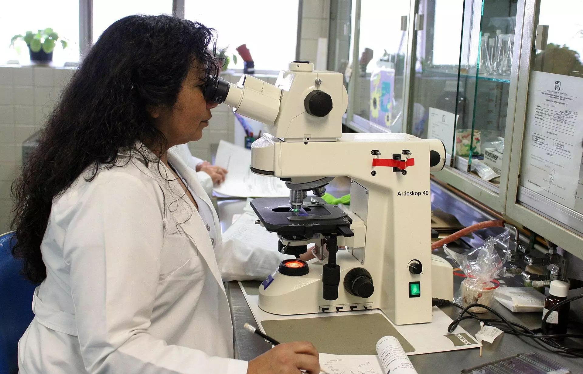
When using a microscope to view biological samples, one issue that arises is the disturbance of the light beam due to the difference in refractive indices between the lens of the objective and the sample. This results in the light rays bending more sharply in the medium surrounding the lens, causing the measured depth in the sample to appear smaller than the actual depth. As a result, the sample may appear flattened when viewed under the microscope.
The Evolution of Corrective Theories
Over the years, various theories have been developed to determine a corrective factor for this depth distortion. However, many of these theories assumed that the corrective factor was constant regardless of the depth of the sample. It wasn’t until the 90s when Nobel laureate Stefan Hell suggested that this scaling factor could be depth-dependent. Sergey Loginov, a former postdoc at Delft University of Technology, has now proven through calculations and a mathematical model that the sample does indeed appear more flattened closer to the lens than farther away.
Following Loginov’s calculations, Ph.D. candidate Daan Boltje and postdoc Ernest van der Wee conducted experiments in the lab to confirm that the corrective factor is depth-dependent. Their findings have been published in the journal Optica. Ernest Van der Wee emphasizes the significance of their research by developing a web tool and software for determining the precise corrective factor for experiments. With these tools, researchers can now accurately adjust for the depth distortion when viewing biological samples under a microscope.
One of the key applications of this research is in protein analysis using electron microscopy. Daan Boltje highlights the importance of precise depth determination in cutting out proteins from biological systems. By accurately determining the structure of proteins, researchers can better understand abnormalities and diseases, leading to potential therapeutic advancements. With the depth-dependent scaling factor, researchers can save time and resources by focusing only on relevant proteins and biological structures.
The web tool provided allows researchers to input the necessary details of their experiment, such as refractive indices, aperture angle of the objective, and light wavelength used. The tool then generates a curve for the depth-dependent scaling factor, which can be exported for further analysis. Researchers can also compare the results with existing theories to gain a better understanding of the depth distortion in their samples.
The discovery of depth-dependent scaling factors in microscopy has revolutionized the way researchers view and analyze biological samples. By accurately adjusting for the depth distortion, researchers can delve deeper into the structures of proteins and biological systems, leading to advancements in understanding and treating diseases. The development of tools such as the web tool and software provided in this research paves the way for more precise and efficient microscopy techniques in the future.


Leave a Reply