In a groundbreaking development, a team of researchers at HHMI’s Janelia Research Campus has successfully applied astronomy techniques to the field of life sciences. The team has utilized methods traditionally used in astronomy to unblur images of galaxies located far away, in order to enhance the clarity and sharpness of microscopy images. This innovation offers biologists a more efficient and cost-effective approach to obtaining high-quality images for their research. The results of this pioneering work have been documented in the journal Optica.
Introduction to Adaptive Optics
Astronomers have long been familiar with techniques that enhance the clarity and sharpness of images captured by telescopes, particularly images of distant galaxies. These techniques involve measuring the distortion of light caused by the atmosphere and applying corrections to eliminate aberrations. Drawing inspiration from these methods, microscopists have been working on adapting similar techniques to improve the quality of images obtained from thick biological samples, which also introduce light bending and distortions.
Despite the promise of adaptive optics in microscopy, the existing methods are known to be intricate, costly, and time-consuming, which limits their accessibility to many research laboratories. Recognizing this limitation, the team at HHMI’s Janelia Research Campus set out to explore a different class of techniques known as phase diversity. While widely used in astronomy, phase diversity methods are relatively new to the life sciences but offer a more straightforward and efficient approach to image enhancement.
Implementation of Phase Diversity
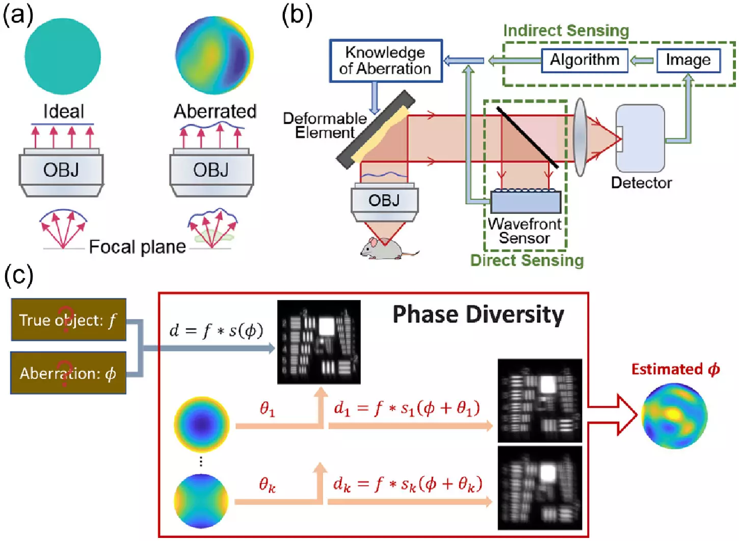
The implementation of phase diversity involves integrating additional images with known aberrations into a blurry image with unknown aberrations. This approach provides the necessary information to deblur the original image effectively. Unlike many other adaptive optics techniques, phase diversity does not necessitate significant modifications to the imaging system, making it an appealing option for microscopy applications. The team at HHMI’s Janelia Research Campus first adapted the astronomy algorithm for microscopy use and validated it through simulations.
To put their new method to the test, the team constructed a microscope equipped with a deformable mirror and two additional lenses, which induce known aberrations. These minor modifications to an existing microscope facilitate the implementation of the phase diversity correction. Through their experiments, the researchers demonstrated a significant improvement in the calibration speed of the microscope’s deformable mirror compared to existing methods. They also showcased the ability of the new technique to detect and correct randomly generated aberrations, resulting in clearer images of fluorescent beads and fixed cells.
Moving forward, the team aims to evaluate the performance of the method on actual biological samples, such as living cells and tissues. Additionally, they plan to extend the utilization of the technique to more sophisticated microscopy systems. The researchers envision further streamlining the method to make it more automated and user-friendly, with the ultimate goal of broadening access to adaptive optics in the biological sciences. The simplified and cost-effective nature of this new method has the potential to revolutionize microscopy practices, enabling researchers to visualize cellular structures with enhanced clarity and precision. Through ongoing advancements and refinements, this innovative approach may pave the way for new discoveries in the field of life sciences.
Leave a Reply