Navigating the intricate landscape of neurological disorders often presents significant challenges, particularly in cases where traditional treatments prove ineffective. Recently, transcranial focused ultrasound (tFUS) has attracted interest as a non-invasive methodology to stimulate targeted brain regions using high-frequency sound waves. This technique boats potential therapeutic applications, especially for individuals suffering from drug-resistant epilepsy and other movement disorders characterized by recurrent tremors.
A research collaboration among experts from Sungkyunkwan University, the Institute for Basic Science, and the Korea Institute of Science and Technology has led to groundbreaking advances in this arena. In an article published in *Nature Electronics*, the team introduced an innovative sensor capable of performing tFUS more effectively than previous models, refining its ability to interact with the brain’s complex topology.
Traditional brain sensors, which are designed to directly contact and monitor the brain surface, have long struggled with accuracy. According to research lead Donghee Son, one of the primary difficulties stems from the sensors’ inability to conform closely to the intricate grooves and folds of the brain. Without this close contact, the capacity to effectively measure neural signals is greatly compromised.
While previous innovations, particularly those developed by Professors John A. Rogers and Dae-Hyeong Kim, provided a degree of improvement by minimizing the physical footprint of the sensors, these devices still faced severe limitations in more curved regions of brain tissue. Their inability to maintain stability due to micro-movements and the natural flow of cerebrospinal fluid greatly hindered their potential clinical applications.
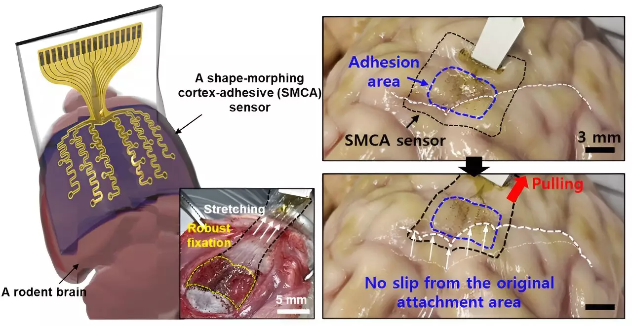
The research team, spearheaded by Son, established a new sensor designed to conquer these challenges. Dubbed ECoG (electrocorticography), this state-of-the-art device merges three crucial layers into its structure, allowing it to adapt and firmly attach to the brain’s convoluted surfaces. This design significantly improves the reliability of ongoing brain signal recordings while utilizing low-intensity focused ultrasound (LIFU) to manage conditions like epilepsy.
Son explained that the ECoG sensor is capable of robust adhesion, eliminating potential voids between the sensor and tissue. This closeness reduces the noise typically associated with external mechanical movements, which is particularly beneficial during ultrasound applications targeting epileptic activity. Such advancements not only optimize the functionality but also increase precision in monitoring brain conditions.
Personalized Treatment Paradigm
The necessity for personalized medical treatment has become more pronounced in contemporary neuroscience. Many efforts are underway to develop individualized ultrasound stimulation therapies, particularly for epilepsy. However, prior to this study, real-time brain wave monitoring often fell short due to the disruptive noise generated from ultrasound vibrations. The noise made it virtually impossible to produce tailored treatment plans that responded to patients’ unique neural activity.
The innovations presented in this latest study aim to change this narrative dramatically. By reducing noise interference and allowing real-time monitoring of brain waves during LIFU, the new sensor facilitates the creation of personalized treatment paradigms. Such capabilities mark a revolutionary step toward enhancing the overall efficacy of neurological treatments.
The ECoG sensor integrates a hydrogel-based layer, a self-healing polymer layer, and a stretchable layer containing gold electrodes. Upon application, the hydrogel layer initiates a rapid bonding process with the brain tissue, which is critical for establishing a strong attachment right from the outset. Following this bond, the self-healing polymer morphs over time to better fit the brain’s contours.
This continuous adaptation significantly enlarges the contact area between the sensor and the brain, allowing for more accurate and consistent measurements—essential for diagnoses and ongoing treatment modifications.
Initial tests on awake rodents yielded promising results, with the ECoG sensor demonstrating its capability to monitor brain waves and manage seizure activity effectively. The research team is not stopping there; plans are underway to upscale the design to create a high-density array of electrodes, thereby enhancing the device’s resolution and diagnostic prowess.
Further development of this technology bodes well not only for epilepsy patients but also shows potential applications in diagnosing and treating various other neurological disorders. As researchers continue to push boundaries, the integration of shape-morphing and tissue-adhesive technologies could pave pathways for enhanced neurological treatments and sophisticated prosthetic solutions.
The development of an innovative transcranial focused ultrasound sensor represents a significant milestone in neurological treatment approaches. With its unique ability to adapt to the brain’s intricate structure, this technology offers promising avenues for personalized medical interventions. As research progresses, it holds the potential to profoundly improve the quality of life for individuals afflicted by challenging neurological disorders.


Leave a Reply