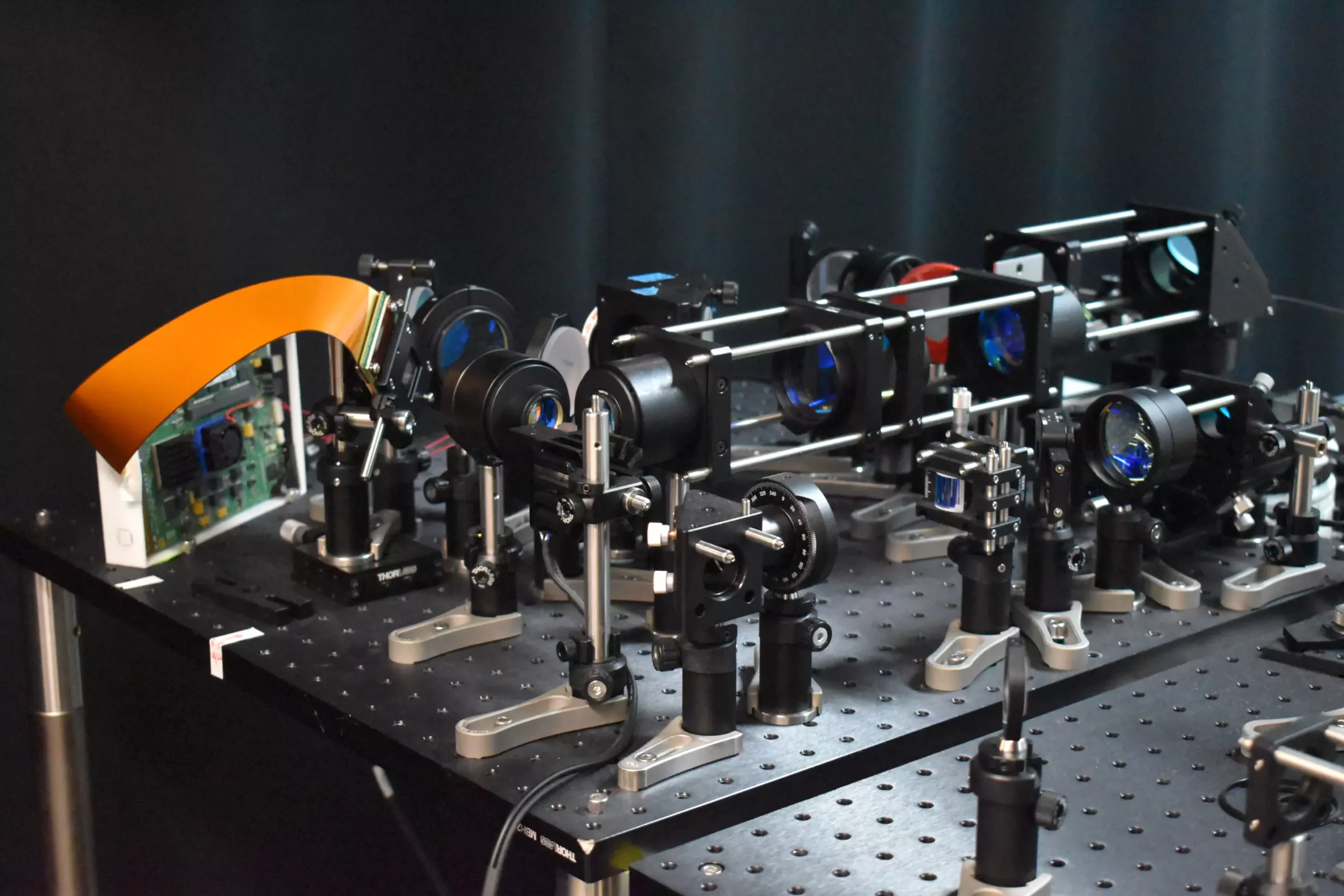
Neuroscientific research has taken a major leap forward with the development of a cutting-edge two-photon fluorescence microscope that is capable of capturing high-speed images of neural activity at cellular resolution. This breakthrough technology offers a significant advancement over traditional two-photon microscopy by enabling faster imaging with minimal damage to brain tissue.
The new microscope, as described in a study published in Optica, incorporates a novel adaptive sampling scheme and replaces conventional point illumination with line illumination. This innovative approach allows for in vivo imaging of neuronal activity in a mouse cortex at speeds ten times faster than previous methods while reducing laser power on the brain by more than tenfold.
Lead researcher, Weijian Yang from the University of California, Davis, emphasizes the importance of real-time imaging in studying the dynamics of neural networks for a better understanding of fundamental brain functions such as learning, memory, and decision-making. By observing neural activity during learning processes, researchers can gain valuable insights into the communication and interaction among different neurons.
The new microscope opens up possibilities for studying the pathology of neurological diseases at their earliest stages. By providing a tool to observe neuronal activity in real time, researchers hope to improve the understanding and treatment of conditions like Alzheimer’s, Parkinson’s, and epilepsy. The ability to monitor rapid neuronal events with high precision could revolutionize the field of neuroscience.
One of the key innovations of the new microscope is the adaptive sampling strategy, which utilizes a line of light to illuminate specific areas of the brain where neurons are active. This not only accelerates the imaging process but also reduces the risk of potential damage to brain tissue. By targeting only the neurons of interest, the total light energy deposited into the brain is minimized.
The incorporation of a digital micromirror device (DMD) in the microscope allows for dynamic shaping and steering of the light beam. This precise targeting of active neurons is crucial for achieving high-resolution imaging and identifying regions of interest quickly. The researchers have demonstrated the ability to isolate the activity of individual neurons, which is essential for understanding complex neural interactions and the functional architecture of the brain.
Looking ahead, the researchers are focused on integrating voltage imaging capabilities into the microscope to capture rapid neural activity directly. This advancement could further enhance the understanding of brain function and lead to new insights into neurological disorders. The goal is to make the microscope more user-friendly and compact, making it more accessible for neuroscience applications.
The development of the new high-speed two-photon fluorescence microscope represents a significant milestone in neuroscience research. By pushing the boundaries of imaging technology, researchers are poised to unravel the complexities of neural activity and pave the way for groundbreaking discoveries in brain science.


Leave a Reply