The use of nonlinear light microscopy has brought about significant advancements in our ability to observe and comprehend complex biological processes. However, one must also consider the potential harm that intense light can inflict on living organisms. Despite this awareness, the exact mechanism behind the irreversible perturbation of cellular processes caused by intense light exposure remains shrouded in mystery.
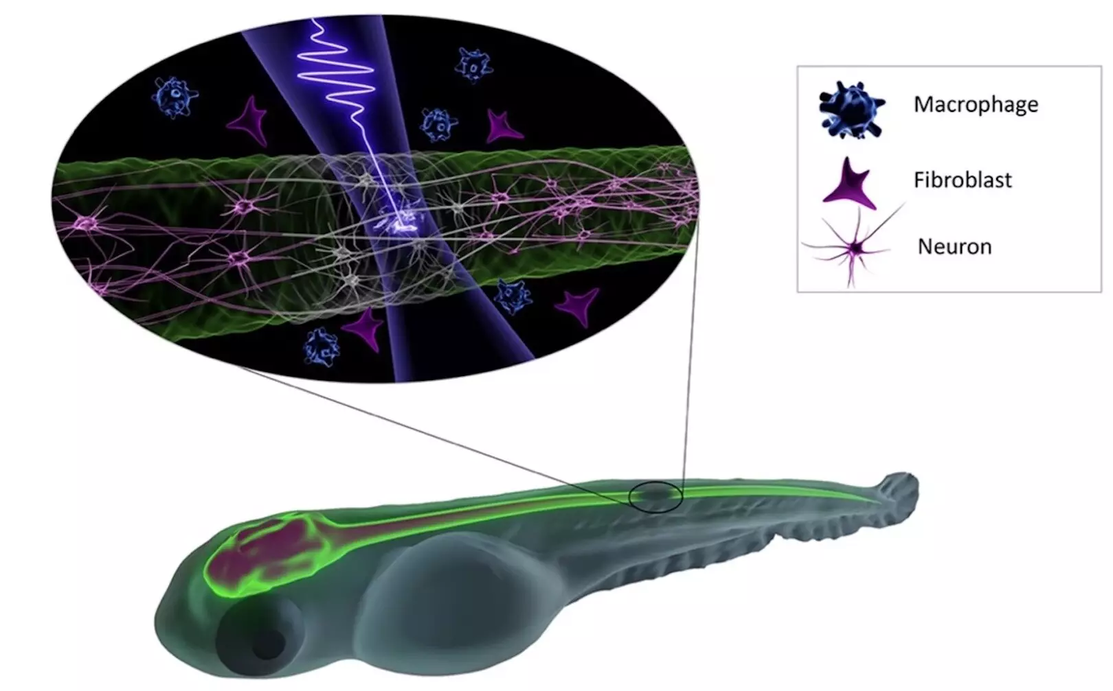
To shed light on this issue, the collaborative research groups of Hanieh Fattahi and Daniel Wehner at the Max Planck Institute for the Science of Light (MPL) and Max-Planck-Zentrum für Physik und Medizin respectively, have embarked on a mission to identify the optimal conditions for using intense pulsed lasers in vivo without causing damage to the organism. By focusing their efforts on the zebrafish, a common vertebrate species used in biological research, the international team was able to investigate the mechanisms of photodamage in deep tissue at a cellular level induced by femtosecond excitation pulses.
The research findings, published in Communications Physics, have shed light on the critical threshold of intense pulsed laser exposure that triggers damage to the central nervous system (CNS) of zebrafish. According to Soyeon Jun, the first author of the publication and a doctoral student in the “Femtosecond Fieldoscopy” group at MPL, the damage occurs abruptly at extreme peak intensities that lead to low-density plasma formation. This discovery opens up new possibilities for noninvasive imaging techniques, allowing for longer imaging dwell times and increased photon flux during irradiation at 1030 nm, as long as the peak intensity remains below the low-plasma density threshold.
The implications of these findings are profound, contributing significantly to the field of deep tissue imaging and innovative microscopy techniques. For instance, femtosecond fieldoscopy, a cutting-edge imaging technique with high spatial resolution and attosecond temporal resolution, is now being developed by Fattahi’s research group. This technique holds promise for label-free imaging and has the potential to revolutionize the way we observe biological processes at the cellular level.
The collaborative efforts between the fields of physics and biology have not only enhanced our understanding of the impact of intense pulsed lasers on deep tissue imaging but also paved the way for future in vivo applications. As noted by Wehner, the head of the research group Neuroregeneration, these findings lay the groundwork for precise light-based manipulations of the central nervous system. This interdisciplinary approach has not only advanced scientific knowledge but also opened up new avenues for research and technological innovation in the field of microscopy and biological imaging.
Leave a Reply